Instruments
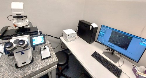
ZEISS Axio Observer
Fluorescence Microscope: ZEISS Axio Observer for confocal-like image quality
- Specification -
- Apotome 3 for optical sectioning in widefield microscopy
- lightsource Colibri 7
- Filter set 90 HE LED
- AI sample finder
- automated x-y-z table, joystick x-y
- objectives:
EC Plan-NEOFLUAR 5x/0,16
EC Plan-NEOFLUAR 10x/0,3
Plan-Apo 20x/0,8
Plan-APO 40x/1,3 Oil DIC (UV)VIS-IR
- b/w and color camera:
Axiocam 705 mono R2
Axiocam 105 color R2
ZEN Software 3.8
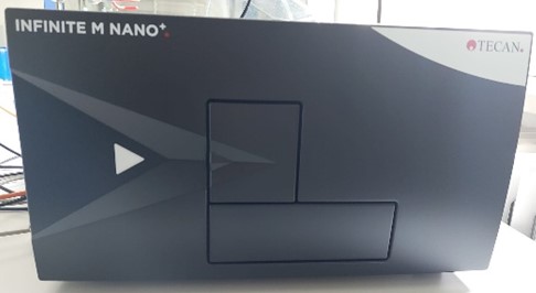
Photometer
Tecan Infinite M Nano+
- Specification -
- Absorbance monochromator + spectral scan 230nm – 1.000nm
- Fluorescence monochromator Ex 230nm – 850nm, Em 280nm – 850nm; top + bottom read
- shake-function
- heating RT +5°C bis 42°C
- i-Control™: Reader Control Software
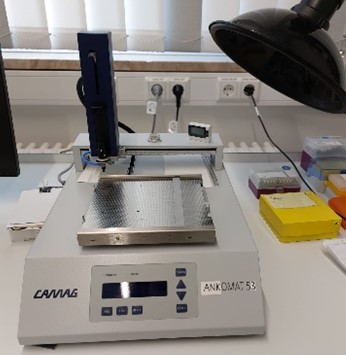
TLC pipetting automate Linomat X

Vibratome Leica VT1200S
- Specification -
- sectioning modes
- automatic
- semi-automatic
- speed
- 0,01 – 0,1 in 0, 01mm/s steps,
- 0,10 – 0,5 in 0, 02mm/s steps,
- 0,50 – 1,5 in 0,1 0mm/s steps
- amplitude
- 0 - 3 mm in 0,05 mm Schritten
- z-axis step size 1, 10 and 100µm (20.000µm uppermost position)
- LED light
- Vibrocheck
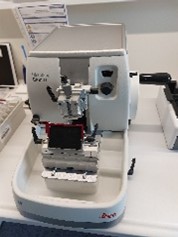
Microtome Leica HistoCore BIOCUT
- Specification -
- manual
- section thickness setting range 0.50 - 100 µm
- trimming thickness setting range 1-600µm
- horizontal feed range 24mm+/-1mm
- vertical stoke length 70+/-1mm
- max cutting range w/o retraction 65mm
- max sectioning area with retraction 60mm
- max specimen block size 55x50x30mm (large standard)
- max specimen block size 68x48x15mm (super cassette)
Molecular biological methods

Thin Layer Chromatography (TLC) was employed to separate and identify lipid classes within complex lipid mixtures, such as sebum. Further studies including the final identification of specific lipid classes and of individual fatty acids are carried via mass spectrometry and gas chromatography in cooperation with our partners.
Western blotting is performed e.g. to qualitatively confirm the presence and activity of Cas9 in transduced cell lines.
Histological Methods

Classical immunohistochemical staining techniques, such as hematoxylin and eosin (H&E) and horseradish peroxidase (HRP)-based staining, are routinely applied for the comparative characterization of sebaceous glands across various species.
(Immuno)fluorescence-stained samples are imaged to enable both qualitative and quantitative image-based analyses, for example to assess lipid metabolism-related manipulations in sebocytes.